


En pratique, l’électrocardiogramme enregistre une activité cardiaque en mesurant le différentiel électrique entre 2 points 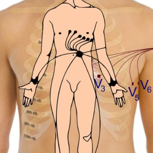 distants sur le coeur.
Tout d’abord, on positionne 10 électrodes, sur la poitrine, sur les bras et sur les jambes. Ensuite, ces électrodes sont reliées à un moniteur qui permet d’observer l’activité du coeur en temps réel.
En principe, cela dure environ 5 minutes et ne requiert aucune préparation particulière.
Ainsi, à l’image d’un sismographe, l’écran affiche une courbe reprenant fidèlement les pulsations cardiaques de votre coeur.
Pour finir, on imprime le tracé de l’électrocardiogramme qui est une sorte de “photographie électrique” de votre coeur.